Surface Topography of Primary Teeth Enamel After Sub-ablative Er;Cr:YSGG Laser Irradiation: An In Vitro Study
Main Article Content
Abstract
Objective: To evaluate the effect of different sub-ablative irradiation parameters of Er;Cr:YSGG laser on the surface topography of primary teeth enamel with white spot lesions.
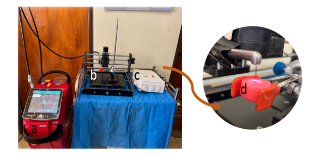
Materials and Methods: A total of 30 primary posterior teeth with sound enamel were immersed in demineralization solution at (pH 4.4) to artificially induce enamel white spot lesions. They were randomly divided into three groups: L1, L2, and L3 groups were irradiated with Er;Cr:YSGG laser irradiation at the power of 0.75W, 0.5W, and 0.25W respectively, 20Hz frequency, and 40% air/ 60% water irrigation. Surface topography was evaluated with a profilometer and scanning electron microscope.
Results: Surface roughness evaluation with scanning electron microscope images revealed a non-significant increase in surface roughness after the demineralization process. Laser irradiation with different powers leads to a non-significant increased surface roughness with altered topography and a more pronounced effect with the laser group L1 and to a lower extent groups L2 and L3.
Conclusion: Increased surface roughness of the primary teeth enamel after sub-ablative power irradiation with Er;Cr:YSGG laser, with a rough and irregular surface devoid of smear layer, the roughness increase was proportional with the increased irradiation power.
Received 24 Apr. 2024; Revised 25 Aug. 2024; Accepted 27 Aug. 2024; Published online 15 Dec. 2024
Corresponding Author:[email protected]
Article Details
Issue
Section

This work is licensed under a Creative Commons Attribution 4.0 International License.


